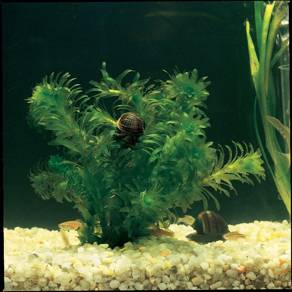
Why Are Cells in the Elodea Plant So Easy to See
Observing Plant Cells

Carolina Labsheets™
In this lab students observe Elodea leaves under magnification. They will see cell walls and chloroplasts. From the movement of chloroplasts they will infer that cyclosis, or protoplasmic streaming, is occurring. They also will observe that most chloroplasts are pressed tightly against the cell wall and should infer from this that much of the cell is occupied by a vacuole.
Needed Materials
Elodea (162101, 162111, or 157340)
springwater (132450) or conditioned tap water
small tanks or large beakers for Elodea
forceps (623990)
dropper(s) (158981)
microscopes (590960)
microscope slides (631920)
coverslips (632900)
dissecting needles (627220)
Optional Materials
In Elodea, cyclosis is easy to observe because chloroplasts move with the cytoplasm as it flows. Light and heat stimulate cyclosis in Elodea. Tungsten or halogen substage microscope lamps produce both heat and light, so after 2–3 minutes, students should be able to observe the movement of chloroplasts. If your microscopes have fluorescent or LED lamps, these produce very little heat and often will not stimulate cyclosis. To provide the needed heat, use a desk lamp equipped with a halogen bulb. Position the lamp so that it shines down on the lab bench. After a few minutes, the surface of the lab bench should become noticeably warm to the touch. Students can place their slides on this warm surface for 3 minutes and then look for signs of cyclosis. A somewhat better arrangement is to position the lamp so that it shines directly onto the stage of a microscope, thereby heating the slide while students view it. Not all slides will show cyclosis, so have students share those that do, so that everyone has the opportunity see the movement.
Safety
Ensure that students understand and adhere to safe laboratory practices when performing any activity in the classroom or lab. Demonstrate the protocol for correctly using the instruments and materials necessary to complete the activities, and emphasize the importance of proper usage. Use personal protective equipment such as safety glasses or goggles, gloves, and aprons when appropriate. Model proper laboratory safety practices for your students and require them to adhere to all laboratory safety rules.
Procedures
To insure that your Elodea is in prime condition for this and other labs, refer to our Elodea Carolina™ CareSheet (available at www.carolina.com). Although you can receive Elodea and use it the same day, it is much better to condition the plants under lights for 2 days.
On the day of the lab set up 2 or more small tanks or large beakers, each containing water and Elodea. Place forceps and droppers alongside each container. Also, set up stations for pickup of microscope slides, coverslips, and dissecting needles.
Optional: If students have studied osmosis, they can observe plasmolysis. Make a 6% solution of NaCl by dissolving 6 g of NaCl in 70 mL of distilled or deionized water and bringing the final volume to 100 mL. (A 6% NaCl solution is approximately 1 M NaCl.)
Students are unlikely to observe nuclei in Elodea cells. However, nuclei are easily observed in stained cells of onion skin. Quarter an onion and separate the layers. Use forceps to remove the skin from the inner (concave) surface of a layer. Cut or tear the onion skin into small pieces that will fit under a coverslip. Place the onion skin on a microscope slide and smooth out as many wrinkles as possible. Add a drop of stain to cover the onion skin. A number of stains can be used, including iodine solutions (iodine-potassium iodide, Lugol solution, Gram iodine), crystal violet, toluidine blue, and methylene blue. After 1 minute, rinse away the stain with tap water, add a coverslip, and observe the cells. Nuclei will be evident.
In addition to light and heat, pH also influences cyclosis. Have students conduct a study to determine at what pH cyclosis is most likely to occur.
Answer Key to Questions Asked on the Student LabSheet
- How many layers of cells do you observe?
Most Elodea leaves have 3 layers of cells. - Do you find that most of the chloroplasts are concentrated against the inner cell wall? If so, what is the likely explanation?
The central part of the cell is filled with a large vacuole.
Note: Under high power magnification students may actually see that the cytoplasm is a thin layer appressed to the cell wall. They may also observe individual chloroplasts as they appear to squeeze through this layer. - The cytoplasm in a cell may move, carrying organelles along with it. Cytoplasmic streaming is called cyclosis. Do you see any indication of cyclosis in the Elodea cells? If so, describe what you see.
Yes. The chloroplasts are moving, indicating that the cytoplasm is moving. - Based on your observations, list at least 2 features of Elodea cells that you would not expect to find in an animal cell.
A cell wall and chloroplasts are not found in animal cells. Students also might infer the presence of a large central vacuole. (Although not directly observed, this is compatible with observations that were made.) - a) If the surface area of an Elodea leaf is 30 mm2 and the average surface area of its cells is 0.0013 mm2 , how many cells compose the leaf? (Remember to account for the cell layers in the leaf.) Show your work.

b) If each cell averages 30 chloroplasts, how many chloroplasts are in the leaf?

Source: https://www.carolina.com/teacher-resources/Interactive/observing-plant-cells/tr38030.tr